Evaluation of Physical, Thermal and Spectral Parameters of Biofield Energy Treated Methylsulfonylmethane
Received: 23-Sep-2015 / Accepted Date: 09-Oct-2015 / Published Date: 19-Oct-2015 DOI: 10.4172/2329-9053.1000129
Abstract
The methylsulfonylmethane (MSM) is an organosulfur compound having sulfonyl functional group. It is occurred naturally in some primitive plants and used in disease related to chronic pain, inflammation, and arthritis. This study was attempted to evaluate the impact of biofield energy treatment on the physical, thermal, and spectral properties of MSM. The study was performed in two groups viz. the control group was remained as untreated, while the treated group was subjected to Mr. Trivedi’s biofield energy treatment. After that, both the control and treated samples were analyzed using surface area analyzer, X-ray diffraction (XRD), thermogravimetric analysisderivative thermogravimetry (TGA-DTG), differential scanning calorimetry (DSC), and Fourier transform infrared (FTIR) spectroscopy. The surface area analysis exhibited a significant decrease in the surface area of treated sample by 22.96% as compared to the control. The XRD analysis showed the significant increase in average crystallite size by 49.20% in the treated sample with respect to the control. The DSC analysis showed the significant increase (67.20%) in latent heat of fusion of treated sample with respect to the control. The TGA analysis showed the onset temperature of thermal degradation at 170°C in the control sample that was slightly decreased to 168.05°C after biofield treatment. Moreover, the Tmax (maximum thermal degradation temperature) was also decreased slightly from 186.66°C (control) to 183.38°C (treated). This indicated the early phase of vaporization in treated sample as compared to the control. The FT-IR spectroscopic study exhibited the alteration in wavenumber of S=O group that suggests the effect of biofield treatment on force constant and bond strength of MSM molecules.
Altogether, the surface area, XRD, thermal analysis and FT-IR spectroscopy suggests that Mr. Trivedi’s biofield energy treatment has the impact on physical, thermal, and spectral properties of MSM.
Keywords: Biofield energy; Methylsulfonylmethane; Surface area; X-ray diffraction; Differential scanning calorimetry; Fourier transform infrared
Abbreviations
NCCAM: National Center for Complementary and Alternative Medicine; NIH: National Institute of Health; XRD: X-Ray Diffraction; DSC: Differential Scanning Calorimetry; TGA: Thermo Gravimetric Analysis; DTG: Derivative Thermo Gravimetry; FT-IR: Fourier Transform Infrared
Introduction
Methylsulfonylmethane (MSM) is a naturally occurring sulfur compound with the formula (CH3)2SO2 [1]. The sulfur content of MSM is helpful to maintain the normal connective tissues in human. It showed the anti-atherosclerotic, anti-inflammatory and free radical scavenging activities [2,3]. As an anti-inflammatory agent, it is used in joint inflammation, osteoarthritis, rheumatoid arthritis, osteoporosis, eye inflammation, chronic pain, muscle cramps, musculoskeletal pain, stretch marks etc. [4,5]. MSM has effectively delayed the tumor onset in colon cancer and mammary breast cancer in rats [6,7]. Moreover, due to its polarity and thermal stability, MSM is used as a high-temperature solvent for wide variety of compounds including polymers organics, and inorganic salts [1]. It is also used as a medium in several organic syntheses like substitution of aryl chlorides with potassium fluoride can be conducted in the presence of MSM as a solvent [8]. By considering the therapeutic importance of MSM and as a solvent in organic/ inorganic synthesis, it is useful to discover an alternate approach, which can improve the physical, thermal, and spectral properties of MSM.
Healing therapy or therapeutic touch is being used as an alternative treatment approach in several fields, and known as the biofield therapy. The National Institute of Health/National Center for Complementary and Alternative Medicine (NIH/NCCAM) considered the biofield energy (putative energy fields) treatment in the subcategory of energy therapies [9]. The biofield energy treatment is used in the healing practice to diminish the anxiety, pain, and to stimulate the overall health of human being [10,11]. The biofield (bioenergetic field) referred to subtle energy field that permeates and surrounds the human body [12]. The health of living organism depends on the balance of this bioenergetics field. In the diseased condition, this bioenergetics field is depleted [13]. The practitioners of energy medicine manipulate and balance this bioenergetics field via harnessing the energy from the Universe [14]. Thus, the human has the ability to harness the energy from the Universe and transfer it to the object to balance or re-pattern the electromagnetic energy field [15]. The objects always receive this energy and respond to the useful way [16]. Biofield energy therapy includes various modalities such as healing touch, therapeutic touch, Reiki, etc. [17]. Mr. Trivedi is well known to possess a unique biofield energy treatment (The Trivedi Effect®) that has been studied in several fields like materials science [18-20], agricultural research [21], biotechnology research [22], microbiology research [23,24], and pharmaceutical sciences [16,25].
Based on the significant impact of biofield energy treatment and chemical importance of MSM, this study was aimed to evaluate the effect of Mr. Trivedi’s biofield energy treatment on physicochemical and spectroscopic properties of MSM. The control and treated samples were analyzed using surface area analysis, X-ray diffraction (XRD), thermogravimetric analysis-derivative thermogravimetry (TGADTG), differential scanning calorimetry (DSC), and Fourier transform infrared (FT-IR) spectroscopy.
Materials And Methods
Study design
The methylsulfonylmethane (MSM) was purchased from S D Fine Chem Limited, India. The MSM was divided into two groups; one was remained as untreated (control), while another group was coded as treated. The treated group in sealed pack was handed over to Mr. Trivedi for biofield energy treatment under laboratory conditions. Mr. Trivedi rendered the biofield energy treatment to the treated group through his unique energy transmission process without touching the sample. Subsequently, the control and treated samples of MSM were evaluated utilizing the several analytical techniques like surface area analyzer, XRD, TGA-DTG, DSC, and FT-IR spectroscopy.
Surface area analysis
The surface area of control and treated MSM was determined using the Brunauer–Emmett–Teller (BET) surface area analyzer (Smart SORB 90) based on the ASTM D 5604 method. The range of the instrument is 0.2 m2/g to 1000 m2/g. Percent change in surface area was calculated with the help of following equation:
Here, SControl is the surface area of the control sample and STreated is the surface area of treated sample.
XRD study
The XRD analysis of control and treated MSM was performed on Phillips, Holland PW 1710 X-ray diffractometer with nickel filter and copper anode. The wavelength of XRD system was set to 1.54056Å. The percentage change in average crystallite size (G) was calculated using following equation:
G=[(Gt-Gc)/Gc] ×100
Here, Gc and Gt are average crystallite size of control and treated powder samples, respectively.
TGA-DTG analysis
The TGA-DTG analysis was performed to determine the effect of biofield treatment on the thermal property of MSM. The analysis was done using Mettler Toledo simultaneous TGA-DTG system. Both the control and treated samples of MSM were heated up to 400°C from room temperature with a heating rate of 5°C/min under air atmosphere. The onset temperature (at which thermal degradation started) and Tmax (temperature at which maximum weight loss occur) in samples were acquired from TGA-DTG thermogram.
DSC study
The melting temperature and latent heat of fusion of control and treated MSM were determined using the Pyris-6 Perkin Elmer differential scanning calorimeter. The samples were heated at the rate of 10°C/min under air atmosphere with air flow rate of 5 mL/min.
FT-IR spectroscopic characterization
The FT-IR spectroscopic analysis was carried out to investigate the impact of biofield energy treatment at the molecular level like dipole moment, force constant, and bond strength in chemical structure [26]. The treated sample was divided into two groups i.e., T1 and T2 based on two different time period (i.e., approximately one month); each sample was then mixed and crushed with spectroscopic grade KBr into fine powder. Finally, the mixture was pressed into pellets and used for FT-IR analysis. The spectra were recorded on Shimadzu’s Fourier transform infrared spectrometer (Japan) with the frequency range of 500-4000 cm-1.
Results and Discussion
Surface area analysis
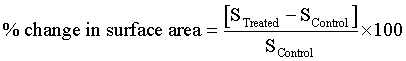
Surface area of both the control and treated samples of MSM was analyzed using BET surface area analyzer and data are shown in Figure 1. The surface area of the control and treated sample was found as 0.3127 m2/g and 0.2409 m2/g, respectively. It showed a significant decrease in surface area by 22.96% in the treated sample as compared to the control. It is well-reported that surface area is inversely proportional to the particle size [27]. Based on this, it was assumed that biofield energy treatment possibly induced the disappearance of inter-particle boundaries and lead to agglomeration in MSM molecule [28]. This may lead to increase in particle size of treated MSM as compared to the control.
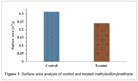
Figure 1: Surface area analysis of control and treated methylsulfonylmethane.
XRD analysis
The XRD diffractograms of control and treated MSM are shown in Figure 2. The control sample exhibited the XRD peaks at 2θ equal to 16.41°, 20.38°, 24.25°, 25.33°, 29.38°, 33.06°, and 50.51°. Similarly, the XRD diffractogram of treated MSM showed the XRD peaks at 2θ equal to 16.47°, 20.40°, 20.51°, 24.16°, 24.32°, 25.32°, and 43.19°. XRD diffractogram showed the intense peaks in both control and treated samples of MSM, which suggest the crystalline nature of samples. Figure 2 showed the significant decrease in the intensity of XRD peaks after biofield treatment with respect to the control sample. Moreover, the most intense peak in control sample was found at 16.41° that was shifted to 20.40° in treated sample. The previously published literature suggests that the alteration in crystal morphology might cause the alteration in relative intensities of the peaks [29]. Furthermore, it is reported that due to presence of internal strain, the 2θ values might change [30]. Based on this it is presumed that biofield energy treatment may increase in internal strain in the treated sample, which might be responsible for alteration in 2θ values of treated sample with respect to control.
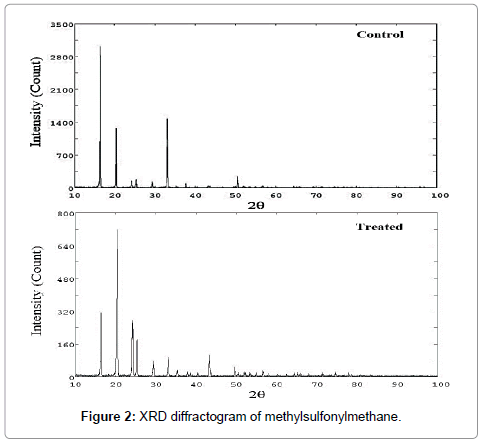
Figure 2: XRD diffractogram of methylsulfonylmethane.
The average crystallite size of the control MSM was observed as 97.76 nm that was increased to 145.86 nm in the treated sample. The result suggests a significant increase (49.2%) in average crystallite size of treated sample as compared to the control (Figure 3). Literature report suggests that increase in annealing temperature significantly affects the average crystallite size of the materials. The increase in temperature leads to decrease in dislocation density and increase in number of unit cell, which finally leads to increase in average crystallite size [31,32].
Figure 3: Average crystallite size of control and treated methylsulfonylmethane.
Based on this, it is assumed that biofield treatment might provide some thermal energy to MSM molecules. Therefore, the dislocation density might be reduced and accordingly the number of unit cell and average crystallite size were increased. The increase in crystallite size might also be correlated with decrease in the surface area of treated sample with respect to the control.
TGA-DTG analysis
The TGA and DTG thermogram of MSM (control and treated samples) are shown in Figure 4 and data are presented in Table 1. The TGA thermogram of control sample showed the initiation (onset) of thermal degradation at 170°C that was terminated (end-set) at 218°C; whereas, in TGA thermogram of the treated sample it appeared at 168.05 and 209.5°C, respectively. The result showed a slight decrease in the onset and end-set temperature of thermal degradation as 1.15 and 3.9%, respectively after biofield energy treatment as compared to the control. The percentage weight loss throughout the thermal decomposition was 52.71% in the control and 53.99% in the treated sample. The result showed a slight increase in percent weight loss during thermal decomposition of treated sample as compared to the control. Overall, the result suggests that biofield treated MSM might be less thermally stable as compared to the control. Furthermore, the DTG thermogram exhibited the Tmax (temperature at which the sample lost its maximum weight) at 186.66°C in the control sample and at 183.38°C in the treated sample of MSM. The result showed about 1.76% decrease in Tmax of treated sample as compared to the control. This might be due to the alteration in internal energy through biofield energy treatment that results into early phase of vaporization in treated sample as compared to the control [33].
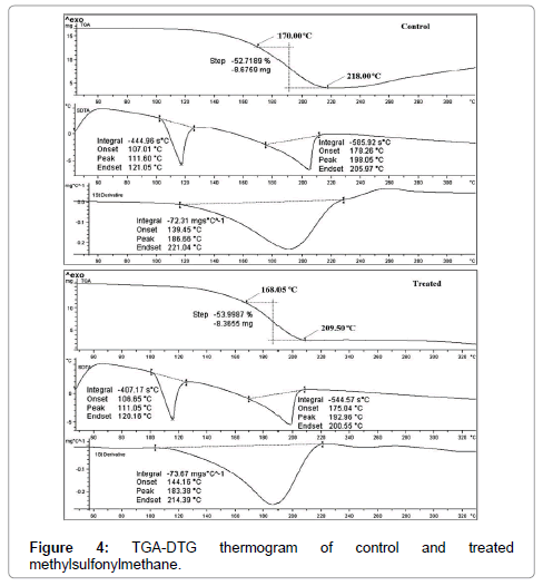
Figure 4:TGA-DTG thermogram of control and treated methylsulfonylmethane.
| Parameter | Control | Treated |
|---|---|---|
| Onset temperature (°C) | 170.00 | 168.05 |
| End-set temperature (°C) | 218.00 | 209.50 |
| Tmax (°C) | 186.66 | 183.38 |
| Latent heat of fusion (J/g) | 97.88 | 163.66 |
| Melting point (°C) | 111.56 | 111.83 |
Table 1: Thermal analysis of control and treated samples of methylsulfonylmethane.
DSC analysis
DSC analysis was done to find out the melting temperature and latent heat of fusion (ΔH) of the MSM (control and treated samples). The DSC thermograms of both samples are shown in Figure 5. It showed the melting temperature at 111.56°C and 111.83°C in the control and treated sample, respectively (Table 1). The result suggests no change in melting temperature of treated sample as compared to the control. The melting temperature of control MSM was well supported by literature data [34]. The latent heat of fusion was observed as 97.88 J/g in control and 163.66 J/g in the treated sample of MSM. The result showed a significant (67.2%) increase in latent heat of fusion of treated MSM as compared to the control. It is well known that melting point of any material is related to its kinetic energy [35]. Based on this, it is expected that biofield treatment may enhanced the internal energy of treated sample as compared to the control and thus led to increase the latent heat of fusion.
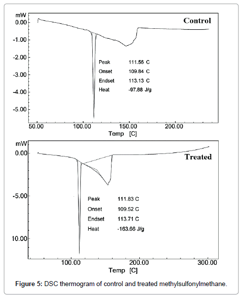
Figure 5: DSC thermogram of control and treated methylsulfonylmethane.
FT-IR spectroscopic analysis
FT-IR spectra of the control and treated samples of MSM are shown in Figure 6. The FT-IR spectra were interpreted with the help of theoretically predicted wavenumber. The MSM molecule contains C-H, C-S, and S=O vibrations. The C-H (methyl) stretching was attributed to peaks at 2933-3016 cm-1 in control and treated (T1) sample, while in T2 sample it was observed at 2933-3018 cm-1. The C-H asymmetrical bending peak was observed at 1425 cm-1 (control and T1) and 1427 cm-1 (T2). The S=O asymmetrical stretching peak was appeared at 1290 cm-1 in control sample, 1288 cm-1 in T1, and 1257 cm-1 in T2 sample. While the S=O symmetrical stretching was observed at 1134 cm-1 (control), 1128 cm-1 (T1), and 1122 cm-1 (T2) sample. The result showed the downstream shifting of S=O asymmetrical (from 1288 cm-1 → 1257 cm-1) and symmetrical stretching (from 1134 cm-1 → 1122 cm-1) peak in treated (T2) sample as compared to the control. This could be due to decreased force constant and bond strength of S=O group in the treated sample [25,26,36]. Therefore, change in frequency might be due to the energy provided via. biofield treatment. According to Sohn et al., change in temperature showed alteration in bond strength of S=O group. Moreover, the C-S stretching was found at 698-761 cm-1 in control sample, 698-763 cm-1 in T1 sample and 700-758 cm-1 in treated sample. Altogether, the FT-IR result showed a significant change in the wavenumber of S=O asymmetrical and symmetrical stretching. The rest of functional groups did not show any significant change in the wavenumber with respect to the control sample.
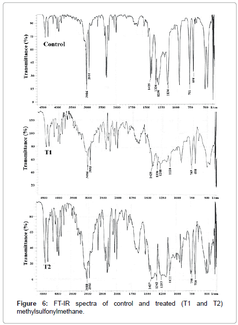
Figure 6: FT-IR spectra of control and treated (T1 and T2) methylsulfonylmethane.
Conclusions
In summary, the result of present study showed the decrease in surface area of treated sample with respect to the control. This might be due to increase in crystallite size of treated sample as evidenced by XRD analysis. The TGA-DTG study showed the slight decrease in onset and end-set temperature of thermal degradation. The DSC analysis exhibited the significant increase in latent heat of fusion of treated sample as compared to the control. This may be due to the increased intermolecular bonding of MSM molecule. The effect of biofield energy treatment on MSM molecule showed time dependent shifting of S=O stretching frequencies in FT-IR spectra with respect to the control. This might be due to the decrease in force constant and atomic bond strength of MSM molecule.
Overall, the present study concluded the significant impact of Mr. Trivedi’s biofield energy treatment on physical, thermal and spectroscopic properties of MSM with respect to the control. Based on this, it is expected that by altering the physicochemical properties using Mr. Trivedi’s biofield energy treatment, the MSM can be changed into the more effective form with respect to its pharmacological activity as well as solvent in organic synthesis.
Acknowledgements
The authors like to acknowledge the Trivedi Science, Trivedi Master Wellness and Trivedi Testimonials for their steady support during the work. Authors would also like to thanks the whole team from the MGV pharmacy college, Nasik fors providing the instrumental facility.
References
- Tapramaz R, Turkkan E, Dereli O (2011) Experimental and theoretical electron paramagnetic resonance (EPR) study on the temperature-dependent structural changes of methylsulfanylmethane. Int J Mol Sci 12: 4909-4922.
- Kim LS, Axelrod LJ, Howard P, Buratovich N, Waters RF (2006) Efficacy of methylsulfonylmethane (MSM) in osteoarthritis pain of the knee: A pilot clinical trial. Osteoarthr Cartil 14: 286-294.
- Beilke MA, Collins-Lech C, Sohnle PG (1987) Effects of dimethyl sulfoxide on the oxidative function of human neutrophils. J Lab Clin Med 110: 91-96.
- Usha PR, Naidu MU (2004) Randomised, double-blind, parallel, placebo-controlled study of oral glucosamine, methylsulfonylmethane and their combination in osteoarthritis. Clin Drug Investig 24: 353-363.
- Castori M (2012) Ehlers-danlos syndrome, hypermobility type: An underdiagnosed hereditary connective tissue disorder with mucocutaneous, articular, and systemic manifestations. ISRN Dermatol 2012: 751-768.
- O'Dwyer PJ, McCabe DP, Sickle-Santanello BJ, Woltering EA, Clausen K, et al. (1988) Use of polar solvents in chemoprevention of 1,2-dimethylhydrazine-induced colon cancer. Cancer 62: 944-948.
- McCabe D, O'Dwyer P, Sickle-Santanello B, Woltering E, Abou-Issa H, et al. (1986) Polar solvents in the chemoprevention of dimethylbenzanthracene-induced rat mammary cancer. Arch Surg 121: 1455-1459.
- Hareau G, Kocienski P (2001) e-EROS Encyclopedia of reagents for organic synthesis. John Wiley & Sons, Ltd. UK.
- Koithan M (2009) Introducing complementary and alternative therapies. J Nurse Pract 5: 18-20.
- Cahil M (1998) Nurses handbook of complementary and alternative therapies. Springhouse, PA: Springhouse Corporation.
- Aldridge D (1991) Spirituality, healing and medicine. Br J Gen Pract 41: 425-427.
- Wilson CA (2011) Healing power beyond medicine. John Hunt Publishing Ltd., UK.
- Warber SL, Cornelio D, Straughn J, Kile G (2004) Biofield energy healing from the inside. J Altern Complement Med 10: 1107-1113.
- Umbreit AW (2000) Healing touch: Applications in the acute care setting. AACN Clin Issues 11: 105-119.
- Trivedi MK, Patil S, Shettigar H, Bairwa K, Jana S (2015) Effect of biofield treatment on spectral properties of paracetamol and piroxicam. Chem Sci J 6: 98.
- Jain S, Mills PJ (2010) Biofield therapies: Helpful or full of hype? A best evidence synthesis. Int J Behav Med 17: 1-16.
- Trivedi MK, Patil S, Tallapragada RM (2013) Effect of bio field treatment on the physical and thermal characteristics of silicon, tin and lead powders. J Material Sci Eng 2: 125.
- Trivedi MK, Patil S, Tallapragada RMR (2015) Effect of biofield treatment on the physical and thermal characteristics of aluminium powders. Ind Eng Manage 4: 151.
- Trivedi MK, Nayak G, Patil S, Tallapragada RM, Latiyal O (2015) Studies of the atomic and crystalline characteristics of ceramic oxide nano powders after biofield treatment. Ind Eng Manage 4: 161.
- Lenssen AW (2013) Biofield and fungicide seed treatment influences on soybean productivity, seed quality and weed community. Agricultural Journal 8: 138-143.
- Nayak G, Altekar N (2015) Effect of biofield treatment on plant growth and adaptation. J Environ Health Sci 1: 1-9.
- Trivedi MK, Patil S, Shettigar H, Gangwar M, Jana S (2015) Antimicrobial sensitivity pattern of Pseudomonas fluorescens after biofield treatment. J Infect Dis Ther 3: 222.
- Trivedi MK, Patil S, Shettigar H, Gangwar M, Jana S (2015) An effect of biofield treatment on Multidrug-resistant Burkholderia cepacia: A multihost pathogen. J Trop Dis 3: 167.
- Trivedi MK, Patil S, Shettigar H, Bairwa K, Jana S (2015) Spectroscopic characterization of chloramphenicol and tetracycline: An impact of biofield. Pharm Anal Acta 6: 395.
- Pavia DL, Lampman GM, Kriz GS (2001) Introduction to spectroscopy. (3rdedn), Thomson Learning, Singapore.
- Groza JR, Shackelford JF (2007) Materials processing handbook. Taylor and Francis group, CRC Press.
- Shih WY, Liu J, Shih WH, Aksay IA (1991) Aggregation of colloidal particles with a finite interparticle attraction energy. J Stat Phys 62: 961-984.
- Inoue M, Hirasawa I (2013) The relationship between crystal morphology and XRD peak intensity on CaSO4.2H2O. J Cryst Growth 380: 169-175.
- Fultz B, Howe JM (2002) In Transmission electron microscopy and diffractometry of materials. Diffraction and the X-ray powder diffractometer. (4thedn), Springer-Verlag: Berlin.
- Raj KJA, Viswanathan B (2009) Effect of surface area, pore volume, particle size of P25 titania on the phase transformation of anatase to rutile. Indian J Chem 48A: 1378-1382.
- Gaber A, Abdel-Rahim MA, Abdel-Latief AY, Abdel-Salam MN (2014) Influence of calcination temperature on the structure and porosity of nanocrystalline SnO2 synthesized by a conventional precipitation method. Int J Electrochem Sci 9: 81-95.
- Sa J (2014) Fuel Production with Heterogeneous Catalysis., CRC Press, Taylor and Francis group LLC., FL, USA.
- Stuart BH (2004) Infrared Spectroscopy: Fundamentals and applications (analytical techniques in the sciences (AnTs). John Wiley & Sons Ltd., Chichester, UK.
Citation: Trivedi MK, Branton A, Trivedi D, Nayak G, Bairwa K, et al. (2015) Evaluation of Physical, Thermal and Spectral Parameters of Biofield Energy Treated Methylsulfonylmethane. J Mol Pharm Org Process Res 3: 129. Doi: 10.4172/2329-9053.1000129
Copyright: ©2015 Trivedi MK, et al. This is an open-access article distributed under the terms of the Creative Commons Attribution License, which permits unrestricted use, distribution, and reproduction in any medium, provided the original author and source are credited.
Share This Article
Open Access Journals
Article Tools
Article Usage
- Total views: 16746
- [From(publication date): 12-2015 - Apr 19, 2024]
- Breakdown by view type
- HTML page views: 12267
- PDF downloads: 4479